
Dog Pythiosis
Canine pythiosis presents as cutaneous or subcutaneous lesions, acquired through open wounds on the skin, or gastrointestinal lesions which are acquired when P. insidiosum contacts the mucous membranes. The fact that dogs frequently drink stagnant water and eat grass that may contain elements of P. insidiosum explains the high number of intestinal pythiosis cases in canines.

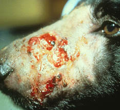
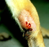
The cutaneous and subcutaneous lesions are denuded of hair and perforated by fistulous sinus tracts that discharge a serosanguineous exudate. Lesions are often located on the dog’s tail, legs, thorax, and abdomen. The hard stony masses (kunkers) observed in equine pythiosis are not present in dogs with this disease. Microscopic examination shows multifocal areas of necrosis with eosinophils and a moderate number of neutrophils and macrophages.
The cutaneous and subcutaneous lesions are denuded of hair and perforated by fistulous sinus tracts that discharge a serosanguineous exudate. Lesions are often located on the dog’s tail, legs, thorax, and abdomen. The hard stony masses (kunkers) observed in equine pythiosis are not present in dogs with this disease. Microscopic examination shows multifocal areas of necrosis with eosinophils and a moderate number of neutrophils and macrophages. The hyphae of P. insidiosum are found in the center of eosinophilic micro abscesses.
Canine gastrointestinal pythiosis is characterized by vomiting, weight loss, and sporadic diarrhea. Formation of hard gastrointestinal tumor-like masses and areas of thickness and mucosal ulceration are common. The infection may spread to adjacent tissue such as pancreas, uterus, and mesenteric lymph nodes. Histopathologically, the mucosa shows ulceration, atrophy, and hyperplasia. Eosinophils, plasma cells, macrophages, epithelial cells and giant cells are detected in infected tissues. The hyphae of P. insidiosum, however, are difficult to detect. Silver stain or other special stains are required to visualize the hyphae of this pathogen in the infected tissues.
DIAGNOSIS: Because P. insidiosum lesions progress rapidly a quick diagnosis is essential for animal survival. Serologic tests employing ELISA technology have been shown to provide both sensitive and specific diagnosis of Pythium infection in dogs.
The hyphae of P. insidiosum are found in the center of eosinophilic micro abscesses.
Canine gastrointestinal pythiosis is characterized by vomiting, weight loss, and sporadic diarrhea. Formation of hard gastrointestinal tumor-like masses and areas of thickness and mucosal ulceration are common. The infection may spread to adjacent tissue such as pancreas, uterus, and mesenteric lymph nodes. Histopathologically, the mucosa shows ulceration, atrophy, and hyperplasia. Eosinophils, plasma cells, macrophages, epithelial cells and giant cells are detected in infected tissues. The hyphae of P. insidiosum, however, are difficult to detect. Silver stain or other special stains are required to visualize the hyphae of this pathogen in the infected tissues.
DIAGNOSIS: Because P. insidiosum lesions progress rapidly a quick diagnosis is essential for animal survival. Serologic tests employing ELISA technology have been shown to provide both sensitive and specific diagnosis of Pythium infection in dogs.
TREATMENT: Because canine pythiosis is a newly described disease, many small animal veterinarians are not familiar with its clinical features. Most cases of cutaneous pythiosis are initially diagnosed and treated as bacterial, parasitic or fungal infections. Gastrointestinal pythiosis is often misdiagnosed as intestinal tumor-like disease (neoplasia) and treated by surgical removal of the tumor mass. Neither of these treatment strategies alone has demonstrated a high level of success.
TREATMENT: Because canine pythiosis is a newly described disease, many small animal veterinarians are not familiar with its clinical features. Most cases of cutaneous pythiosis are initially diagnosed and treated as bacterial, parasitic or fungal infections. Gastrointestinal pythiosis is often misdiagnosed as intestinal tumor-like disease (neoplasia) and treated by surgical removal of the tumor mass. Neither of these treatment strategies alone has demonstrated a high level of success.
SURGERY: Once the diagnosis of pythiosis have been established, wide surgical removal of the infected tissues may be a successful treatment for cutaneous, subcutaneous, and intestinal pythiosis. However if the P. insidiosum is not completely removed the lesions will re establish.
DRUGS: Antifungal therapy using amphotericin B, itraconazole, ketoconazole, or nistatin has been unsuccessful.
IMMUNOTHERAPY: Treatment with the therapeutic product used to cure cases of equine pythiosis has shown 35% success in dogs with pythiosis. This may be due in part that all treated cases were dogs with chronic pythiosis (infection >60 days). Immunotherapy has demonstrated a lower rate of cure in chronic infections. This immunotherapeutic product has show greater than 75% success in treating infections of less than 30 days duration in Equines. 6/16/04