
Horse Pythiosis
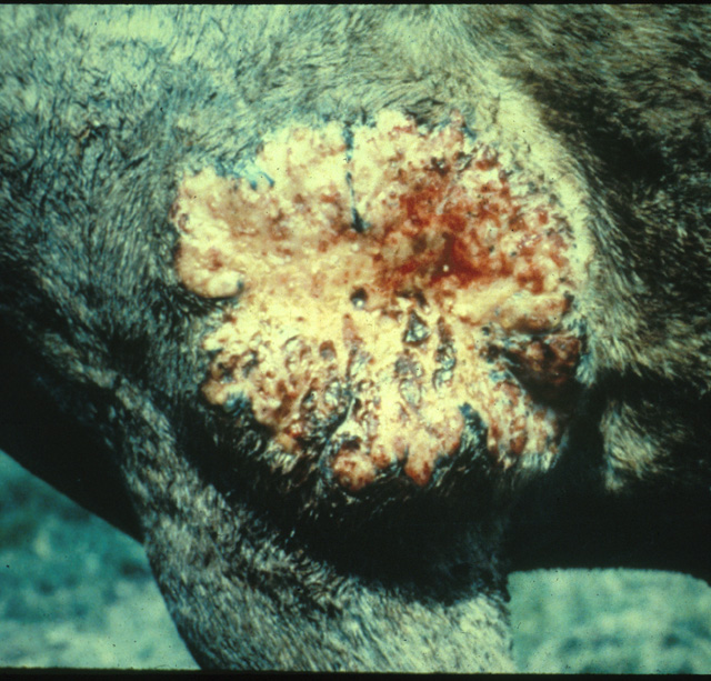
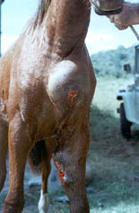
Equine pythiosis is characterized by the development of cutaneous, subcutaneous, lymphatic and intestinal lesions and less frequently by the involvement of bones and lungs (chronic pythiosis). Lesions caused by P. insidiosum can occur on any part of the horse’s body. Lesions of the lower limbs are more common due to more frequent contact with the organism in infested environments (stagnant water, grasses). The lesions often occur singly, but cases with multiple granulomatous lesions have been encountered. There are no reports of animal to animal, or animal to human transmission of this pathogen. If the disease is not treated in the early stages it is fatal in >95% of cases. In most cases treatment with antifungal drugs is not helpful.

Lesions on the limbs are characterized by the formation of tumor like masses with fistulas and a serosanguineous discharge. Lesions on the thorax, abdomen, and shoulders, tend to be circular, 5 to 500 mm in diameter. Ulceration and pruritus (itching) is commonly associated with large lesions. The formation of small hard coral-like masses termed “kunkers” is an interesting characteristic of the disease in equines. These stony masses contain the viable hyphae of P. insidiosum surrounded by cell detritus from degranulated eosinophils.
Lesions on the limbs are characterized by the formation of tumor like masses with fistulas and a serosanguineous discharge. Lesions on the thorax, abdomen, and shoulders, tend to be circular, 5 to 500 mm in diameter. Ulceration and pruritus (itching) is commonly associated with large lesions. The formation of small hard coral-like masses termed “kunkers” is an interesting characteristic of the disease in equines. These stony masses contain the viable hyphae of P. insidiosum surrounded by cell detritus from degranulated eosinophils.
Metastasis (spread) from distant lesions, through lymphatic vessels to regional lymph nodes, lungs, or bones have been reported. Like dog pythiosis, intestinal equine pythiosis is more likely to be acquired by direct inoculation of the organism through ingestion, than spread from distant lesions.
Histopathologically, in early equine pythiosis, abundant micro abscesses with eosinophils, a few neutrophils, lymphocytes, and macrophages are present. In chronic cases, an eosinophilic granuloma with giant cells is observed. In the center of the micro abscesses, stony masses (kunkers) are often present. With Periodic Acid Schiff (PAS) and Silver stains P. insidiosum appears as sparsely septate hyphae 6 to 10 mm in diameter.
TREATMENT:
SURGERY — The most common treatment of equine pythiosis has been the surgical removal of the lesions. This method is very popular and frequently used by veterinary practitioners. A common short-coming of surgical treatment is its high rate of recurrence. This is due to the incomplete removal of the P. insidiosum from the affected tissues. Additionally, surgical recession of lesions of the limbs is very difficult to accomplish without permanent damage to the surrounding tissues.
CHEMOTHERAPY — Two main groups of Antimycotic drugs have been used to treat pythiosis: Iodine and amphotericin B. Both drugs, however, have given contradictory results.
TREATMENT:
SURGERY — The most common treatment of equine pythiosis has been the surgical removal of the lesions. This method is very popular and frequently used by veterinary practitioners. A common short-coming of surgical treatment is its high rate of recurrence. This is due to the incomplete removal of the P. insidiosum from the affected tissues. Additionally, surgical recession of lesions of the limbs is very difficult to accomplish without permanent damage to the surrounding tissues.
CHEMOTHERAPY — Two main groups of Antimycotic drugs have been used to treat pythiosis: Iodine and amphotericin B. Both drugs, however, have given contradictory results. For instance, some practitioners reported that iodine can cure the disease after intravenous injections while others reported failures with the same procedures. In theory, amphotericin B should not work on P. insidiosum due the fact that this pathogen does not have ergosterol (target of the drug) in its cytoplasmic membrane. Nevertheless, the drug has been used with some success in equine pythiosis. The use of drugs in treating pythiosis has been limited because of cost, poor success rate, and high toxicity.
IMMUNOTHERAPY — In the early 1980’s an immunotherapeutic therapeutic product for treatment of P. insidiosum infections in equines was developed in Costa Rica. This early immunotherapeutic product cured 100% of the acute cases (infection 60 days). It was found that horses with chronic infections often become immunodepressed due to the loss of large quantities of proteins, electrolytes and water through the open wounds. Thus, the immunotherapeutic product works better in equine with intact immune system (early pythiosis). A new formulation of this therapeutic vaccine has been introduced by Michigan State University and Pan American Veterinary Laboratories. This new formulation cured 50% of the chronic cases that the original immunotherapeutic product failed to cure. The overall (acute plus chronic) rate of cure of this new immunotherapeutic product was 75%.

For instance, some practitioners reported that iodine can cure the disease after intravenous injections while others reported failures with the same procedures. In theory, amphotericin B should not work on P. insidiosum due the fact that this pathogen does not have ergosterol (target of the drug) in its cytoplasmic membrane. Nevertheless, the drug has been used with some success in equine pythiosis. The use of drugs in treating pythiosis has been limited because of cost, poor success rate, and high toxicity.
IMMUNOTHERAPY — In the early 1980’s an immunotherapeutic therapeutic product for treatment of P. insidiosum infections in equines was developed in Costa Rica. This early immunotherapeutic product cured 100% of the acute cases (infection 60 days). It was found that horses with chronic infections often become immunodepressed due to the loss of large quantities of proteins, electrolytes and water through the open wounds. Thus, the immunotherapeutic product works better in equine with intact immune system (early pythiosis). A new formulation of this therapeutic vaccine has been introduced by Michigan State University and Pan American Veterinary Laboratories. This new formulation cured 50% of the chronic cases that the original immunotherapeutic product failed to cure. The overall (acute plus chronic) rate of cure of this new immunotherapeutic product was 75%.