
Diagnosis of Pythiosis
The early diagnosis of pythiosis is of importance for its successful treatment. Several methodologies have been used for its diagnosis: wet mount preparations, culture, histopathology, and serology.
Wet Mounts: The collected tissue from the infected areas is sent to the laboratory in sterile distilled water at room temperature. Pieces of the tissue are placed with 10% KOH. The finding of sparsely septate hyphae may be indicative of P. insidiosum, or other fungal pathogen (zygomycetes).

Culture: To isolate this organism is important to remember that P. insidiosum is severely inhibited by low temperatures. Thus, transportation of the biopsy tissue in ice will decrease the chance to isolate this pathogen in culture. Samples, therefore, should be sent to the laboratory in sterile water at room temperature. Small pieces of the biopsy
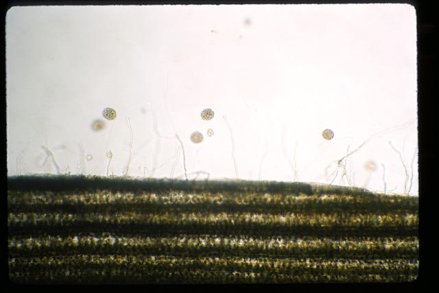
Culture: To isolate this organism is important to remember that P. insidiosum is severely inhibited by low temperatures. Thus, transportation of the biopsy tissue in ice will decrease the chance to isolate this pathogen in culture. Samples, therefore, should be sent to the laboratory in sterile water at room temperature. Small pieces of the biopsy tissue should be placed onto Sabouraud dextrose agar plates and incubated at 37C, the ideal temperature for P. insidiosum primary isolation. Pythium insidiosum grows rapidly at 37C, but incubation at room temperature delays its growth rate. Small colonies should be observed around the tissue sample after 24 to 48 hours at Sporangia developing on grass 37C. At 5 days the plate will be filled with submerged filamentous fungi-like growth. Microscopically, hyphae without sporulation are observed. To identify P. insidiosum the formation of zoospores must be induced in water cultures with, grass leaves, with specific ionic strength. Sporangia containing zoospores (asexual stage) will be observed at edges of the grass. The production of oogonium (sexual stage) is rare.
Histopathology: Tissue sections of the biopsy samples in H&E stains show a typical eosinophilic inflammatory reaction. The hyphae of P. insidiosum, however, are difficult to observe with this stain. Silver stain and Periodic Acid-Schiff (PAS) are suggested for the proper identification of the hyphal elements of P. insidiosum in tissue.
Serology: Several serologic tests have been developed to diagnose pythiosis in humans and animals.
tissue should be placed onto Sabouraud dextrose agar plates and incubated at 37C, the ideal temperature for P. insidiosum primary isolation. Pythium insidiosum grows rapidly at 37C, but incubation at room temperature delays its growth rate. Small colonies should be observed around the tissue sample after 24 to 48 hours at Sporangia developing on grass 37C. At 5 days the plate will be filled with submerged filamentous fungi-like growth. Microscopically, hyphae without sporulation are observed. To identify P. insidiosum the formation of zoospores must be induced in water cultures with, grass leaves, with specific ionic strength. Sporangia containing zoospores (asexual stage) will be observed at edges of the grass. The production of oogonium (sexual stage) is rare.
Histopathology: Tissue sections of the biopsy samples in H&E stains show a typical eosinophilic inflammatory reaction. The hyphae of P. insidiosum, however, are difficult to observe with this stain. Silver stain and Periodic Acid-Schiff (PAS) are suggested for the proper identification of the hyphal elements of P. insidiosum in tissue.
Serology: Several serologic tests have been developed to diagnose pythiosis in humans and animals.
Complement fixation for the diagnosis of equine pythiosis was developed in Australia. It is a sensitive test but lacked specificity. This test is no longer in use in the laboratories dealing with P. insidiosum.
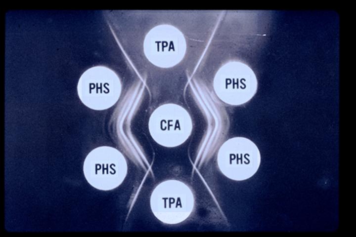
Immunodiffusion (ID) has been the most widely employed serological test to diagnose pythiosis in
Complement fixation for the diagnosis of equine pythiosis was developed in Australia. It is a sensitive test but lacked specificity. This test is no longer in use in the laboratories dealing with P. insidiosum.
Immunodiffusion (ID) has been the most widely employed serological test to diagnose pythiosis in humans and animals. The test is very specific but it has demonstrated a low level of sensitivity. The finding that the sera from some humans and dogs with proven pythiosis gave negative results indicated that the diagnosis using ID has to be confirmed with a more sensitive test.

humans and animals. The test is very specific but it has demonstrated a low level of sensitivity. The finding that the sera from some humans and dogs with proven pythiosis gave negative results indicated that the diagnosis using ID has to be confirmed with a more sensitive test.

Enzyme-Linked Immunosorbent Assay (ELISA) was developed to specifically detect the hyphae of P. insidiosum in the biopsy tissue from humans and animals. The technique uses peroxidase labeled polyclonal antibodies against P. insidiosum to bind to the hyphae in infected tissue. The tissue sections are examined microscopically to visually identify the stained hyphae.
Immunoperoxidase assay was developed to specifically detect the hyphae of P. insidiosum in the biopsy tissue from humans and animals. The technique uses peroxidase labeled polyclonal antibodies against P. insidiosum to bind to the hyphae in infected tissue. The tissue sections are examined microscopically to visually identify the stained hyphae.
Fluorescent antibody technique was developed to diagnosis pythiosis from fixed tissue samples and to identify P. insidiosum from culture. The technique specifically detected P. insidiosum hyphae and gave negative results when tested against Entomophthorales and Mucorales zygomycetes.
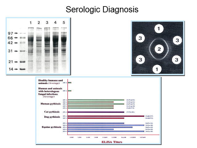
Western blot assay has been used primarily for research purposes. It detects several antigenic proteins of P. insidiosum with sera from patients with Pythiosis. It is both sensitive and specific but is of limited usefulness in clinical diagnosis due to its need for special equipment and its expense.